Journée Futur & Ruptures

Last thursday (January 24th 2013) I participated in the Journée Futur & Ruptures 2013, held at the Institut Mines-Télécom in Paris, France. This nice french name indicates the program of which my PhD project is part.

The goal of the program Futur & Ruptures is to implement a long-term vision and test new approaches on topics such as future telecommunication networks, digital contents, information technology and computer-assisted healthcare.

The aim of this annual meeting is to facilitate the interactions among all PhD students and the parts that financially support and sponsor the program Futur & Ruptures. In this context, each PhD students was invited to present their project in a specific format. In my case, I prepared a poster 🙂

I’ve also updated the Publications Section of my blog. If you are curious to read my poster, just send me an e-mail at this address:  andrea.collo AT lirmm.fr

hammers, screws and Intramedullary nails

If you study orthopaedics, you’ll know for sure that IM femoral nailthe femur is the longest, heaviest and strongest bone in the human body. One day, by chance, it happens that you see a bar like the one in the picture on the right. Well, maybe you’ll also notice two things: it is as long as your femur bone and it has more or less the same curvature. With a bit of fantasy and science fiction mood, you could even get to figure out a strange surgery where such long bars are plugged into the bones of some patients in order to strengthen their skeleton and create an army of solid soldiers.

There’s no need to invent anything like that, since we are talking about a surgery that already exists (but not for military purposes). Intramedullary nailing consists in forcing a metal nail (or rod) into the medullary cavity of a bone. Normally, IntraMedullary (IM) nails are employed to treat fractures of long bones of the body. Nowadays, this surgery is regarded as the standard of treatment for both femoral and tibial shaft fractures.

Let’s focus on an IM femoral nail. What about its surgery?

First, the surgeon makes use of a Reamer. Wikipedia defines it as a “metalworking tool used to create an accurate sized hole“.  The medical version of this device is actually employed to hollow out the center of the medullary canal of the femur, by accessing it from the top (next to the pelvis bone). This doesn’t affect too much the bone solidity and is necessary when the medullary cavity is not continuous (for example, in the case of broken leg injuries).

Once the femur has been properly drilled, an intramedullary nail is tapped into place. This means that a hammer is used to push the rod down into the hollow medullary cavity. In a sense, this action recreates a well-defined medullary cavity in the case of displaced or unstable fractures. Moreover, it obviously provides stabilization for the healing bone (that starts growing again in a proper “shape”).

Finally, screws are usually placed in the head of the femur to secure the nail and prevent its collapse or rotation. During the progressive return to activity, the leg of the patient will take benefit from the fact that the loads will be shared by both the healing bone and its solid metal core. This leads also to a faster rehabilitation period (which requires a first non-weight bearing stage followed by specific exercise programmes).

hammer IM nail

After the implantation, the intramedullary nail is usually left inside the bone forever. In some cases, the patient may develop some long-term complications that cause pain and general ache at the insertion site. In such cases, a second surgery might be necessary to remove the IM nail from the recovered bone. Wikipedia gives us some interesting data about long-term complications of IM nailing for the femur, that “may include persistent or permanent knee pain (present in 73.2% of patients), atrophy of the calf muscle (27.3%), atrophy of the quadriceps (27.3%), and arthritis (35.4%)“.

The following image shows the femoral fracture (A) and the IM nail inserted all along the bone (B), the relatively small scar on the patient’s leg (C) and the recovered bone (D, E).

IM nailing steps

sources: Wiki, Journal of Orthopaedic Science and About.com

Knuckle Popping is addictive

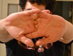
Awesome people always pop their knuckles when they are with someone who clearly state they can’t stand it. The cracking sound that our knuckles (but also our fingers themselves, and sometimes our elbows and knees) produce when we “stretch” them is somehow addictive. I thought it would be interesting to understand what makes our joints pop in this strange way and I asked God Google for that.

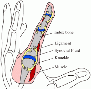
First of all we have to focus on Diarthrodial Joints, better known (maybe) as Synovial Joints. We have synovial joints all over our body: in our hands, wrists, arms, but also in our shoulders and knees. Wikipedia actually says that a synovial joint is “the most common and most movable type of joint in the body of a mammal”.

As probably said in joint capsulesome previous post, in this kind of articulation two bones get in contact with each other through cartilage surfaces. A Joint Capsule acts as a connective tissue that folds the whole articulation and keeps everything at the right place, ensuring mobility and stability at the same time. This capsule is filled by Synovial Fluid, which has two main functions:

  • it continuously lubricates the articulation;
  • it is a source of nutrients for the cells that maintain the joint cartilage.

The synovial fluid contains dissolved gases, mainly oxygen (O2), nitrogen (N2) and carbon dioxide (CO2). These gases are responsible for the popping sound we are trying to explain! After this necessary introduction (yep, I’ve just spoiled the name of the murderer), let’s come back to the action of cracking our knuckles.

crack 1 crack 2In order to pop our knuckles, we can stretch or bend our fingers. In any case, the bones of each knuckle joint (which is a synovial joint) pull apart. As we can easily figure out, by doing this the knuckle joint capsule gets stretched.

knuckleThis causes the volume of the joint capsule to increase a bit (+ 15-20%). This slight change of volume is followed by a corresponding decrease of the synovial fluid pressure. As a consequence, the gases in the fluid suddenly become less soluble and they form small bubbles inside the tiny joint capsule. The process of “rapid pressure change -> formation of small cavities in the liquid -> formation of bubbles that immediately implode” is defined by fluid dynamics as cavitation.

The implosion of such small bubbles is thought to be the origin of the cracking sound that we hear while popping our knuckles. It normally takes 20-30 minutes for the gas to properly redissolve into the synovial fluid and reestablish the initial conditions. This means that after more or less half an hour of silence we’ll be able to start having fun again 🙂

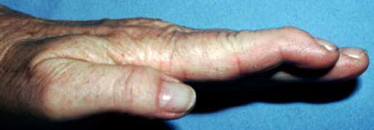
Many people seem to be frightened by the idea that excessive knuckle popping may lead to unpleasant consequences, such as arthritis or sudden death (…). Luckily, a few studies confirm that apparently there is no correlation between knuckle cracking and osteoarthritis in the finger joints. i love itAnother study, however, showed that pathological addiction to knuckle popping may affect the joint capsule soft tissue (higher risk of damages and wear) and worsen hand grip strength conditions. On the positive side, there’s evidence of slightly increased mobility in joints right after popping (mainly thanks to muscle relaxation induced by this action).

In conclusion, we can crack our knuckles whenever we want, but we’d better do it in moderation.

sources: this website, this other one and wikipedia 

Mallet Finger: don’t try this at home

There are a few everyday life experiences that everybody is destined to go through every now and then. Like correctly plugging a USB device only at the third attempt (despite there are only two possibilities), or directly setting the alarm clock half an hour earlier because we know we’re used to putting it off at least four times, or having Mallet Finger.

Mallet Finger is probably one of the most painful and annoying injuries of all time. Technically, it is an injury of the extensor digitorum tendon of the fingers at the distal interphalangeal joint (DIP). In more simple terms, it is the typical injury that occurs when we play basketball and the ball suddenly hits our extended finger. Besides the immediate sensation of pain, within a few minutes our finger will start swelling and we won’t be able to straighten it for a while. We then leave the court with an awesome facial expression (it really hurts, you all know…), but do we know what happened inside our finger?

mallet finger The distal interphalangeal joint (DIP) hinge jointof the hand is nothing more than a hinge joint between the two last phalanges of the finger. This kind of joint only admits one degree of freedom, which is the rotation about the joint axis. As a result, our phalanges are allowed to make flexion and extension movements. Thus, the DIP is the last joint of the finger. A sudden high force acting at the tip of the finger (the ball we were trying to catch) strongly solicits the thin DIP extensor tendon. In case of rupture, or tearing, of this tendon from the bone, the finger usually gets painful, swollen, and bruised. Occasionally, blood can collect beneath the nail. In the worst case, the force of the blow may even pull away a piece of bone along with the tendon. mallet finger bruised The loss of extensor tendon continuity might lead to severe consequences and must be carefully treated. In a first moment, ice should be immediately applied and the hand should be elevated above the level of the heart. Medical attention should be sought within a week after injury. Most mallet finger injuries can be treated without surgery. fingertip splintsNormally, X-rays are necessary in order to look for potential bone fractures or joint misalignment. The presence of blood beneath the nail and nail detachment may be a sign of nail bed laceration or open (compound) fracture. A splint can be applied to hold the fingertip straight (in extension) until it heals (8 weeks full-time, 3-4 further weeks less frequently). With this treatment plan, the finger usually regains an acceptable function and appearance. Despite that, it is not guaranteed that the patient will be able to regain full fingertip extension.

If nonsurgical treatment fails, after mallet finger surgeryconsultation with an orthopaedic surgeon the patient may consider to resort to surgical repair. In case of very severe deformity or inability to properly use the injured finger, surgery is done to repair the fracture using pins, pins and wire, or even small screws. Surgical treatment of the damaged tendon can include tightening the stretched tendon tissue, using tendon grafts, or even fusing the joint straight.

_

sources: mainly this website and this website, and then Google Images