what about goosebumps ?

porcupineThere’s one thing that porcupines will always do better than humans: having goosebumps. Ok, ok, we cannot raise our quills when threatened, simply because we do not have quills. But when we have goosebumps, our body hairs behave exactly in the same way as porcupine quills do. Cutis anserina, a definitely less catchy way to call it, consists in the formation of bumps on the skin. The curious thing is that this phenomenon is involuntary 🙂 let’s try to understand how this happens.

goosebumpsA feeling of cold, a sudden strong emotion of fear, pleasure, euphoria and, yes, also sexual arousal… Our body reacts to all these events in the simplest way possible: trying to protect itself. And we cannot control it, since it’s a reflex (click here to read about another reflex typical of human body). In a previous post we learnt an interesting thing about human body thermoregulation: homeostasic processes (that we saw also here) always try to keep our Body Temperature (BT) of 37°C despite environment conditions. When outside it’s too cold, our energy losses get more important and our BT lowers too fast. We know that, for example, if we do some physical exercise (even a short run) we’ll warm up again quite fast. This is because the activation of muscles develops that energy needed to warm up the body and restore proper BT conditions. But when a sudden feeling of cold occurs, our skin receptors immediately send this information to the brain via the sympathetic nervous system. Our brain cannot wait for us to take a decision and, as previously said, automatically activates a protective action: shivering.
By doing this, our muscles produce really fast contractions that we cannot control (don’t forget we’re always dealing with a reflex!).

arrector pili muscles

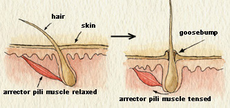
The twitching movements of muscles produce heat, which helps to raise BT. The contraction of the arrector pili muscles, that are the tiny muscles at the base of each hair, pulls the hair erect. In that moment our body acts like that of a porcupine, even if the latter experiences this reflex when threatened (by appearing larger, the animal intimidates enemies).

In exactly the same way, if our jaw muscles begin to shiver, we start chattering our teeth. The mechanism is always the same: BT lowering is detected and an automatic response is activated to raise it up again. In an extremely stressful situation, it is possible to have goosebumps also after experiencing the so-called fight or flight response, when (from this webpage) “the sympathetic nervous system floods the blood with adrenaline (epinephrine), a hormone that speeds up heart rate, metabolism, and body temperature in the presence of extreme stress”. But this is another story that we’ll see later. For tonight, don’t forget to feed your porcupine with a wonderful home-made soup (possibly warm)!

other sources: uno, due e tre

Mallet Finger: don’t try this at home

There are a few everyday life experiences that everybody is destined to go through every now and then. Like correctly plugging a USB device only at the third attempt (despite there are only two possibilities), or directly setting the alarm clock half an hour earlier because we know we’re used to putting it off at least four times, or having Mallet Finger.

Mallet Finger is probably one of the most painful and annoying injuries of all time. Technically, it is an injury of the extensor digitorum tendon of the fingers at the distal interphalangeal joint (DIP). In more simple terms, it is the typical injury that occurs when we play basketball and the ball suddenly hits our extended finger. Besides the immediate sensation of pain, within a few minutes our finger will start swelling and we won’t be able to straighten it for a while. We then leave the court with an awesome facial expression (it really hurts, you all know…), but do we know what happened inside our finger?

mallet finger The distal interphalangeal joint (DIP) hinge jointof the hand is nothing more than a hinge joint between the two last phalanges of the finger. This kind of joint only admits one degree of freedom, which is the rotation about the joint axis. As a result, our phalanges are allowed to make flexion and extension movements. Thus, the DIP is the last joint of the finger. A sudden high force acting at the tip of the finger (the ball we were trying to catch) strongly solicits the thin DIP extensor tendon. In case of rupture, or tearing, of this tendon from the bone, the finger usually gets painful, swollen, and bruised. Occasionally, blood can collect beneath the nail. In the worst case, the force of the blow may even pull away a piece of bone along with the tendon. mallet finger bruised The loss of extensor tendon continuity might lead to severe consequences and must be carefully treated. In a first moment, ice should be immediately applied and the hand should be elevated above the level of the heart. Medical attention should be sought within a week after injury. Most mallet finger injuries can be treated without surgery. fingertip splintsNormally, X-rays are necessary in order to look for potential bone fractures or joint misalignment. The presence of blood beneath the nail and nail detachment may be a sign of nail bed laceration or open (compound) fracture. A splint can be applied to hold the fingertip straight (in extension) until it heals (8 weeks full-time, 3-4 further weeks less frequently). With this treatment plan, the finger usually regains an acceptable function and appearance. Despite that, it is not guaranteed that the patient will be able to regain full fingertip extension.

If nonsurgical treatment fails, after mallet finger surgeryconsultation with an orthopaedic surgeon the patient may consider to resort to surgical repair. In case of very severe deformity or inability to properly use the injured finger, surgery is done to repair the fracture using pins, pins and wire, or even small screws. Surgical treatment of the damaged tendon can include tightening the stretched tendon tissue, using tendon grafts, or even fusing the joint straight.

_

sources: mainly this website and this website, and then Google Images

the Knee Bursae: some hints

The bursae of the knee can be defined in a very simple way: they are fluid sacs, or synovial pockets. This second definition comes from the sinovial fluid that fills them.

Synovial fluid is made of hyaluronic acid and lubricin, proteinases and collagenases. Its main functions are reducing friction by lubricating the joint, absorbing shocks and properly “feeding” joint cartilage. In the case of the knee, the Knee Capsule encloses the Knee Cavity which is filled with synovial fluid. Knee Bursae surround and sometimes communicate with the Knee Cavity, as we can see in the picture.

Usually Knee Bursae are thin-walled and represent the weak point of the joint. At the same time, their presence is really important since they enlarge the joint space. They can be grouped according to:

  • their characterization as communicating and non-communicating bursae. A communicating bursa is when a bursa is located adjacent to a joint, thus having the synovial membrane in communication with the joint itself.
  • their location (frontal, lateral, medial).

In pathological conditions, such as excessive local friction, infection, arthritides or direct trauma, fluid and debris collect within the bursa or fluid extends into the bursa from the adjacent joint. As a consequence, the walls of the bursa thicken as the bursal inflammation becomes longstanding. The term bursitis refers to pathological enlargement of the bursa. Clinically, bursitis mimics several peripheral joint and muscle abnormalities.


<–prepatellar bursitis

          elbow bursitis–>

_

_

sources: Wikipedia and this website

Patellar Reflex

definition _

Patellar Reflex, also called knee-jerk, is a stretch reflex associated with quadriceps femoris muscle stretching.

how it works _

The stretch is created by a blow upon the patellar tendon (positioned just below the Patella). This blow, usually performed with a specific tendon hammer, activates the muscle spindle in the quadriceps femoris muscle.

Muscle spindle is a particular type of sensory receptor, normally embedded in muscle fibers, able to detect changes in the length of the muscle itself. Once “activated” by the external blow, this receptor sends a signal to the spinal cord. Instead of involving higher nervous centres (it would take too long), at the level of the spinal cord an alpha-motor neuron is immediately activated.

The alpha-motor neuron conducts an efferent impulse directly back to the quadriceps femoris muscle, leading to its contraction. At the same time, an inhibitory interneuron relaxes the hamstring muscle, which is the quadricep’s antagonistic muscle.

The result of such coordinated contraction-relaxation, causes the “kick movement” of the leg. In normal health conditions, the leg extends once and then comes back to rest. It only takes about 50 milliseconds between the tap and the start of the leg kick.

why it is useful _

Patellar Reflex is a proprioceptive reflex which helps keeping posture and balance. The fact that everything “happens” at the level of the spinal cord, without involving higher nervous centres, allows for instance to keep balance without effort (actually, one does not have to focus on keeping an upright position). Energies are saved for more complex activities. Moreover, Patellar Reflex helps avoiding strong muscle contractions which could tear the tendon.

clinical interest _

As said, there is no interneuron in the pathway leading to contraction of the quadriceps muscle. Patellar Reflex can be used, for example, to check the conditions of the connections between the spinal cord and the muscles.

The absence or decrease of the Patellar Reflex is known as Westphal’s sign. On the other hand, multiple oscillation of the leg following the blow may be a symptom of cerebellar diseases.

source: contents taken from Wikipedia’s page