Knee Surgery… there’s an app for that!

app

Nowadays “there’s an app for that” could be the 11th Commandment … there is a bunch of apps for everything, even for the most stupid daily activities.

vks1A friend of mine told me to have a look at this one: Virtual Knee Surgery. It is a very realistic game that follows all the basic steps of a Total Knee Replacement surgery. The user is invited to interact with the surgeon in order to perform the necessary operations.

After living this experience in a real operating room, personally I’ve found this app a really nice and clear explanation 🙂 If you want to spend ten minutes to learn the basics of TKR, I definitely can say it’s worth it!

vks2

Mallet Finger: don’t try this at home

There are a few everyday life experiences that everybody is destined to go through every now and then. Like correctly plugging a USB device only at the third attempt (despite there are only two possibilities), or directly setting the alarm clock half an hour earlier because we know we’re used to putting it off at least four times, or having Mallet Finger.

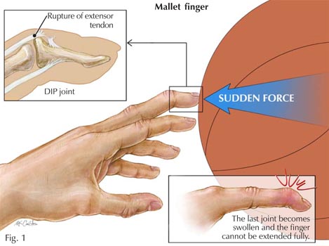
Mallet Finger is probably one of the most painful and annoying injuries of all time. Technically, it is an injury of the extensor digitorum tendon of the fingers at the distal interphalangeal joint (DIP). In more simple terms, it is the typical injury that occurs when we play basketball and the ball suddenly hits our extended finger. Besides the immediate sensation of pain, within a few minutes our finger will start swelling and we won’t be able to straighten it for a while. We then leave the court with an awesome facial expression (it really hurts, you all know…), but do we know what happened inside our finger?

mallet finger The distal interphalangeal joint (DIP) hinge jointof the hand is nothing more than a hinge joint between the two last phalanges of the finger. This kind of joint only admits one degree of freedom, which is the rotation about the joint axis. As a result, our phalanges are allowed to make flexion and extension movements. Thus, the DIP is the last joint of the finger. A sudden high force acting at the tip of the finger (the ball we were trying to catch) strongly solicits the thin DIP extensor tendon. In case of rupture, or tearing, of this tendon from the bone, the finger usually gets painful, swollen, and bruised. Occasionally, blood can collect beneath the nail. In the worst case, the force of the blow may even pull away a piece of bone along with the tendon. mallet finger bruised 
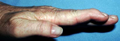
The loss of extensor tendon continuity might lead to severe consequences and must be carefully treated. In a first moment, ice should be immediately applied and the hand should be elevated above the level of the heart. Medical attention should be sought within a week after injury. Most mallet finger injuries can be treated without surgery. fingertip splints
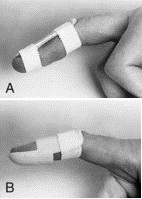
Normally, X-rays are necessary in order to look for potential bone fractures or joint misalignment. The presence of blood beneath the nail and nail detachment may be a sign of nail bed laceration or open (compound) fracture. A splint can be applied to hold the fingertip straight (in extension) until it heals (8 weeks full-time, 3-4 further weeks less frequently). With this treatment plan, the finger usually regains an acceptable function and appearance. Despite that, it is not guaranteed that the patient will be able to regain full fingertip extension.

If nonsurgical treatment fails, after mallet finger surgery
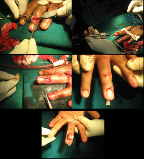
consultation with an orthopaedic surgeon the patient may consider to resort to surgical repair. In case of very severe deformity or inability to properly use the injured finger, surgery is done to repair the fracture using pins, pins and wire, or even small screws. Surgical treatment of the damaged tendon can include tightening the stretched tendon tissue, using tendon grafts, or even fusing the joint straight.

_

sources: mainly this website and this website, and then Google Images

the Ankle Joint: some hints

The ankle is the region where the foot and the leg meet. The ankle joint is actually composed by three smaller joints:

  1. the ankle joint proper, commonly called ankle mortise joint (but also talocrural joint).  It is a synovial hinge joint that connects the distal ends of both the tibia and the fibula in the lower limb with the proximal end of the talus.
  2. the subtalar joint, that occurs at the meeting point of the talus and the calcaneus.
  3. the inferior tibiofibular joint, between the fibula and the tibia. More precisely, it is formed by the rough, convex surface of the medial side of the distal end of the fibula, and a (corresponding) rough concave surface on the lateral side of the tibia.

ankle joint and its three sub-joints

The boney architecture of the ankle consists of three bones: the tibia, the fibula (in the leg) and the talus (in the foot). The talus is also called the ankle bone since it’s the most important bone in the ankle articulation. In normal health conditions, the articulation between the tibia and the talus (ankle mortise joint) bears the greatest part of body weight: it is the region where ankle efforts are mostly concentrated.

The medial malleolus is a boney processmalleoli extending distally off the medial tibia. There is also a lateral malleolus, generated by a distal-most aspect of the fibula. Together, the two malleoli, along with their supporting ligaments, stabilize the talus underneath the tibia.

The ankle joint is bound by the strong deltoid ligament (it is attached at the medial malleolus of the tibia and supports the medial side of the whole joint) and three lateral ligaments: the anterior and posterior talofibular ligaments (they support the lateral side of the joint, from the lateral malleolus to the dorsal and ventral ends of the talus) and the calcaneofibular ligament (it is attached at the lateral malleolus and to the lateral surface of the calcaneus).

ankle ligaments

Achille's tendonThe calcaneus is also attached to the Achille’s tendon (also known as the calcaneal tendon or the tendo calcaneus), that is a tendonous extension of gastrocnemius and soleus muscles of the leg. It attaches the heel to the posterior leg.

Concerning the joint motion, the ankle joint theoretically admits 1 degree of freedom: movements of plantar flexion and dorsiflexion.

ankle motions

In addition to these, the geometry of the different bones that form the articulation permits other more limited movements, such as foot eversion and inversion.

sources: Wikipedia and this website

the Knee Bursae: some hints

The bursae of the knee can be defined in a very simple way: they are fluid sacs, or synovial pockets. This second definition comes from the sinovial fluid that fills them.

Synovial fluid is made of hyaluronic acid and lubricin, proteinases and collagenases. Its main functions are reducing friction by lubricating the joint, absorbing shocks and properly “feeding” joint cartilage. In the case of the knee, the Knee Capsule encloses the Knee Cavity which is filled with synovial fluid. Knee Bursae surround and sometimes communicate with the Knee Cavity, as we can see in the picture.

Usually Knee Bursae are thin-walled and represent the weak point of the joint. At the same time, their presence is really important since they enlarge the joint space. They can be grouped according to:

  • their characterization as communicating and non-communicating bursae. A communicating bursa is when a bursa is located adjacent to a joint, thus having the synovial membrane in communication with the joint itself.
  • their location (frontal, lateral, medial).

In pathological conditions, such as excessive local friction, infection, arthritides or direct trauma, fluid and debris collect within the bursa or fluid extends into the bursa from the adjacent joint. As a consequence, the walls of the bursa thicken as the bursal inflammation becomes longstanding. The term bursitis refers to pathological enlargement of the bursa. Clinically, bursitis mimics several peripheral joint and muscle abnormalities.


<–prepatellar bursitis

          elbow bursitis–>

_

_

sources: Wikipedia and this website

Functional Rehabilitation Robotics: the ‘mPower 1000’ arm brace

The mPower 1000 is a powered arm brace that fits like a sleeve on a person’s arm. The arm brace has myoelectric sensors that sit on the skin’s surface and detect even a very faint muscle signal. When a person with a weak or partially paralyzed arm tries to move their arm and a muscle signal fires, the motor in the mPower 1000 engages to assist in completing the desired movement. In helping achieve desired movement, the device can be worn as a functional aid, used during exercise to maintain gains or applied  during rehabilitation to facilitate Repetitive Task Practice that re-teaches arm movement to the brain.

The mPower 1000 is based on technology developed at MIT, and is lightweight and portable. It has on-board controls for easy use and built-in Bluetooth capability for communication with external applications and systems. The mPower 1000 is for use in the home and in clinical settings. It is intended to increase arm movement for individuals affected by brain injury such as stroke, spinal cord injury (SCI), multiple sclerosis (MS), cerebral palsy (CP), muscular dystrophy (MD) and traumatic brain injury (TBI).

source: this website

what’s LaTIM ?

The Laboratory of Medical Information Processing (LaTIM – INSERM UMR 1101) is a research team made up of the University Hospital of Brest, the Faculty of Medicine and Telecom Bretagne (graduate engineering school). It carries out many projects in three main research areas:

  1. Computer-Assisted Orthopaedic Surgery (IPAL research team);
  2. Quantitative Multimodal Imaging for Diagnosis and Therapy (Quantitative Multi-modality Imaging Team);
  3. Multimedia Medical Information Indexing, Tracking and Integrity (IT2IM Team).

These three themes are integrated within a methodological approach of the diagnostic and therapeutic imaging based on information and knowledge. For my PhD project I am member of the IPAL Team, carrying out my research in the field of Computer-Assisted Orthopaedic Surgery (CAOS).

A day at the hospital

Last week I was invited to attend in a knee surgery at the Hospital Cavale Blanche, in Brest. For my PhD project, the research work that I am currently carrying on is actually closer to the mechanical side of Robotics (microactuators, tiny mechanical structures, wireless energy transfer techniques …). Although, since one of my supervisors is an orthopedic surgeon, I was invited to participate to TKA surgery. Basically, this meant two things: I had to put on specific sterilized clothes and, once in the operating room, I was allowed to watch and put questions 🙂

When I got here I was told that, for PhD students in the field of Robotic Surgery, half a day at the hospital was more useful, in terms of acquirable knowledge, than a month of theoretical study. Actually, one of the aspects I mostly appreciated was the “pedagogical approach” of the surgeon towards the observers. We were two, a sweet girl who’s completing her studies in Pharmacology and me. And the surgeon kept explaining each single step, motivating each procedure and answering all our questions. The confidence he showed while (literally) manipulating the patient’s knee and conducting the operation in front of younger and less experienced surgeons was simply amazing.

Before the surgery, the patient can decide to undergo either general or local anesthesia. Let’s say that the noises and, especially, the smells produced by hammers and saws are not the best way to stimulate one’s appetite 😉 But one thing is sure: after living the atmosphere of such a delicate surgical operation, and understanding that one can really trust expert surgeons, personally I wouldn’t be so worried/scared to undergo TKA as I was before 🙂